- Health
- Mind
- Neuroscience
A new study reveals that nerve cells receive periodic infusions of mitochondria from neighboring cells — and this may point to a new way of treating nerve pain.
When you purchase through links on our site, we may earn an affiliate commission. Here’s how it works.
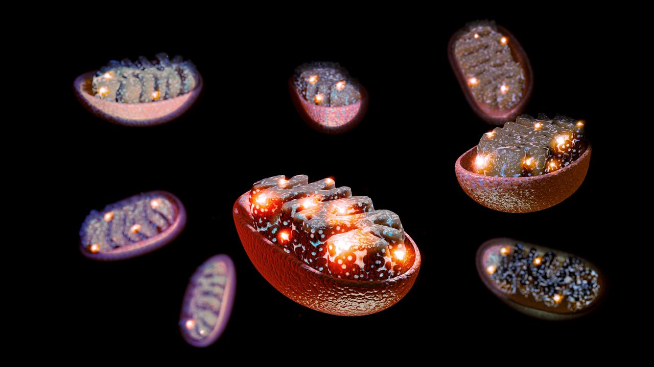
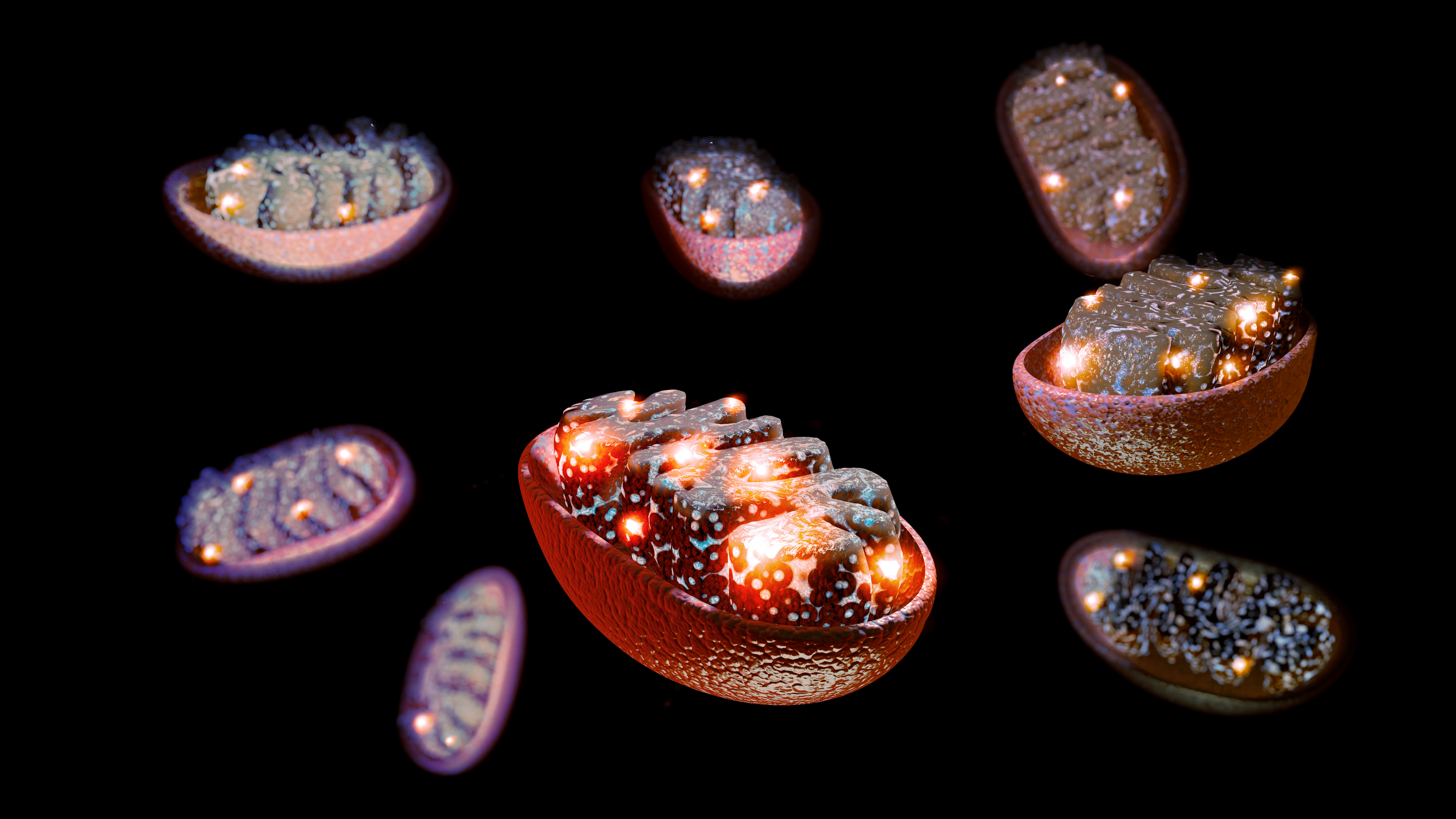 The powerhouses of cells, the mitochondria, may be key to protecting nerves from damage and dysfunction.
(Image credit: Naeblys via Getty Images)
Share
Share by:
The powerhouses of cells, the mitochondria, may be key to protecting nerves from damage and dysfunction.
(Image credit: Naeblys via Getty Images)
Share
Share by:
- Copy link
- X
Supplying nerves with a fresh supply of mitochondria could curb chronic nerve pain, a new study hints.
The research, conducted with mouse cells, live mice, and human tissues, reveals a previously unsung role of mitochondria, the powerhouses of cells. It shows that support cells within the nervous system can ship mitochondria to the nerves that respond to pressure, temperature and pain. But problems with that shipping process can deplete the nerves' energy reserves, causing them to malfunction.
You may like-
 New drug could prevent diabetes complications not fixed with blood sugar control, study hints
-
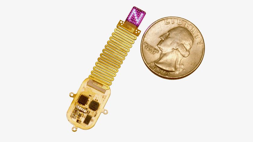 Tiny implant 'speaks' to the brain with LED light
-
 Slaying 'zombie cells' in blood vessels could be key to treating diabetes, early study finds
The new study, published Wednesday (Jan. 7) in the journal Nature, points to potential new ways of heading off that neuronal breakdown — and one strategy could involve transferring mitochondria directly into nerves.
Fresh mitochondria reduce pain
The research zoomed in on satellite glial cells, unique cells that physically wrap themselves around the "roots" of nerve cells located near the spinal cord. The bodies of these nerve cells cluster together near the spine, and from each cluster, bundles of long fibers extend to different parts of the body, from head to toe. The longest of these fiber bundles belong to the sciatic nerve, which measures just over 3 feet (1 meter) long.
The sheer length of the fibers poses a "real challenge," because for a nerve to function properly, mitochondria made in the nerve's root must travel down to the end of each fiber, and that in itself requires energy to do, Ji said. That raises a question of how nerves maintain this power-hungry supply chain.
Sign up for the Live Science daily newsletter nowContact me with news and offers from other Future brandsReceive email from us on behalf of our trusted partners or sponsorsBy submitting your information you agree to the Terms & Conditions and Privacy Policy and are aged 16 or over.Scientists once thought that cells had to make all of their own mitochondria, but in recent years, they have uncovered evidence that cells swap mitochondria. This can occur between cells of the same type or between cells of different types, such as between a stem cell and an immune cell, for example. To facilitate the swap, cells construct tiny structures called tunneling nanotubes for the mitochondria to travel through, like spitballs sliding from one end of a straw to another.
Ji and his team wondered whether satellite glial cells might be able to send mitochondria to the nerve cells they encircle — and it turns out that they can.
"We demonstrate that these cells actually extend these tunneling nanotubes to deliver in the mitochondria. This [finding] is unique in this study," Ji said.
You may like-
New drug could prevent diabetes complications not fixed with blood sugar control, study hints
-
Slaying 'zombie cells' in blood vessels could be key to treating diabetes, early study finds
-
 'Chemo brain' may stem from damage to the brain's drainage system
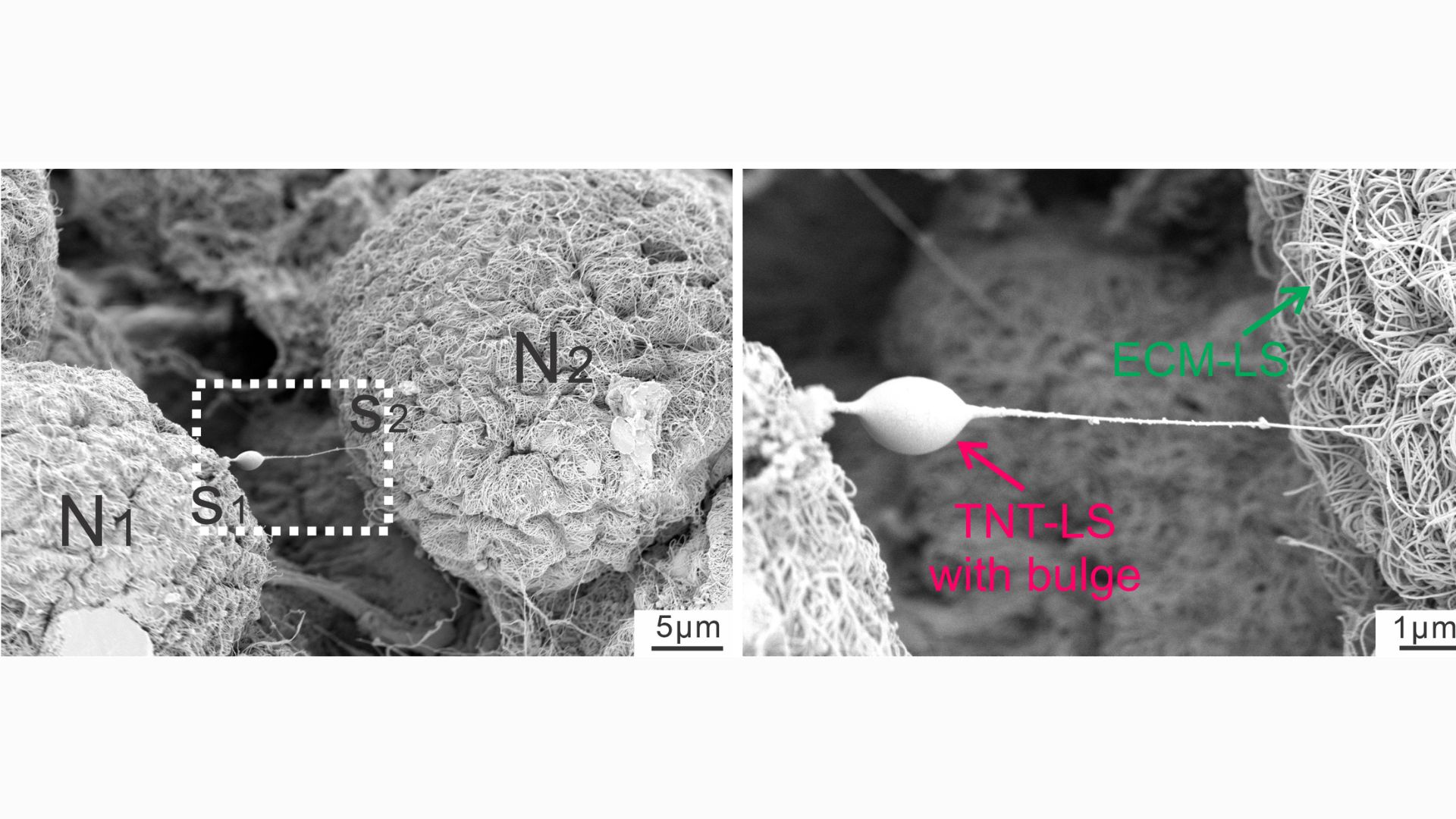 (Image credit: Xu et al. Nature (2026). doi: 10.1038/s41586-025-09896-x)
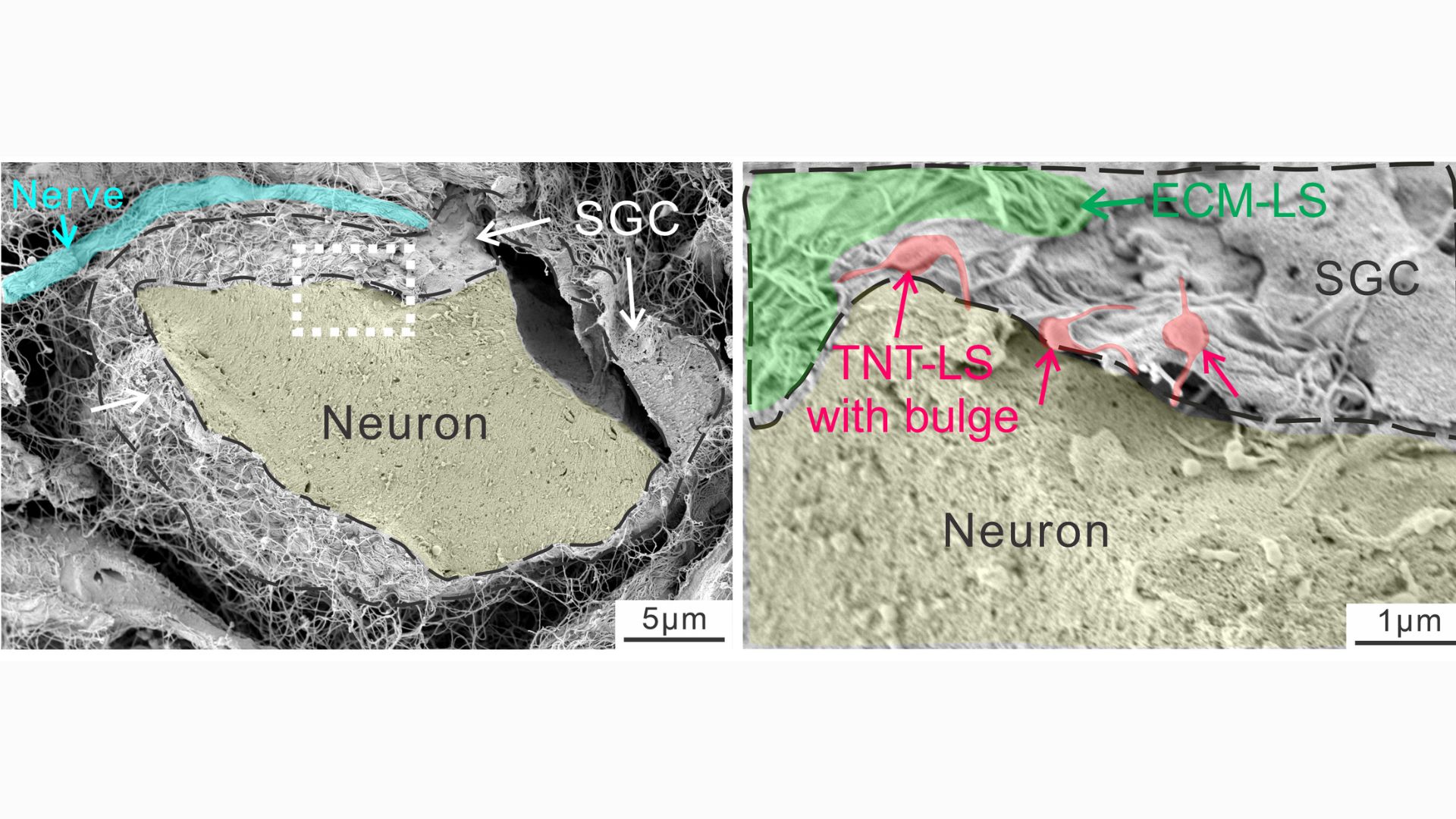
This close-up snapshot shows a nanotube extending from a neuron. The bulge in the tube indicates that something is being transported down its length.
 (Image credit: Xu et al. Nature (2026). doi: 10.1038/s41586-025-09896-x)
This diagram shows nanotubes linking glia to neurons, with bulges in the tubes indicating things being transported from the former cells to the latter. Later analyses revealed these transports to be mitochondria.
In a series of experiments with mouse cells and human tissues, the researchers took snapshots of the tiny tubes that formed between glia and nerve cells, noting distinct "bulges" that appeared in the tubes as materials traveled through them. By tacking a fluorescent tag onto mitochondria, they were able to track instances in which powerhouses from glial cells made their way into the nerves.
The nanotubes were transient structures that broke down soon after a given transfer was complete. Experiments showed that a protein called MYO10 was critical to the tubes' construction, helping to extend them out from the glia. But additionally, the mitochondria could sometimes be transferred without the tubes, either inside tiny bubbles released by glia or through special channels that formed between the membranes of the donor and recipient cells.
In healthy lab mice, the researchers found that disrupting these different modes of mitochondria shipment made the mice more sensitive to pain. That's because it spurred damage in the nerves and caused them to fire abnormally.
They also looked at mice with various types of nerve damage, such as from exposure to chemotherapy drugs or from diabetes. These nerve-damaging conditions also disrupted the mitochondrial exchange from glia to some degree, and this contributed to nerve pain in the lab mice. Transferring healthy glia into the mice alleviated the pain, though, by providing them with a fresh source of healthy mitochondria.
A new view on glia
Notably, nerve damage from diabetes and chemotherapy tends to hit the smallest nerve fibers the hardest, whereas medium and large fibers show more resilience. In the team's experiments, they found that the larger nerve fibers appeared to receive a higher volume of mitochondria from glia, while small fibers got fewer by comparison. In short, it seems that glia have a "preference" toward lending their mitochondria to larger fibers, the study authors wrote.
"That is still a puzzle. We don't know why that's the case," Ji said. But nonetheless, it might begin to explain why small fibers are more vulnerable to damage in these conditions, triggering symptoms of numbness, painful tingling or burning in the feet and hands.
More studies are needed to fully understand how mitochondria are shuttled from glia to nerve cells in health and disease. This fundamental research could pave the way to future treatments for nerve pain, the team thinks. In theory, treatments could be aimed at boosting the activity of satellite glial cells, so they produce and transfer more mitochondria.
Or alternatively, mitochondria could be harvested from cells grown in the lab, purified, and then injected straight into nerves as a treatment, he added.
RELATED STORIES—Tiny implant 'speaks' to the brain with LED light
—New pocket-size model of ALS 'breathes and flows like human tissue'
—Strikingly simple 'dial' in the brain may help it distinguish imagination from reality
Historically, glia were solely thought of as the glue of the nervous system, providing structural support to neurons by binding them together. But scientists have since uncovered that glia are involved in processes once thought to be handled only by neurons, like memory. And the new study suggests glia may actually be physically plugged into neuronal networks, Ji said.
"If they can transport mitochondria, such a very large organelle, in that tube, then you can transport many other things, right?" he suggested. "That means the neurons and the glial cells, they are much more connected than we thought."
 Nicoletta LaneseSocial Links NavigationChannel Editor, Health
Nicoletta Lanese is the health channel editor at Live Science and was previously a news editor and staff writer at the site. She holds a graduate certificate in science communication from UC Santa Cruz and degrees in neuroscience and dance from the University of Florida. Her work has appeared in The Scientist, Science News, the Mercury News, Mongabay and Stanford Medicine Magazine, among other outlets. Based in NYC, she also remains heavily involved in dance and performs in local choreographers' work.
Show More CommentsYou must confirm your public display name before commenting
Please logout and then login again, you will then be prompted to enter your display name.
Logout Read more New drug could prevent diabetes complications not fixed with blood sugar control, study hints
 Slaying 'zombie cells' in blood vessels could be key to treating diabetes, early study finds
 'Chemo brain' may stem from damage to the brain's drainage system
 Gene on the X chromosome may help explain high multiple sclerosis rates in women
 Brain benefits of exercise come from the bloodstream — and they may be transferrable, mouse study finds
 Injecting anesthetic into a 'lazy eye' may correct it, early study suggests
Latest in Neuroscience
Injecting anesthetic into a 'lazy eye' may correct it, early study suggests
Latest in Neuroscience
 Tiny implant 'speaks' to the brain with LED light
 Neuroscience word search — Find all the parts of the brain
 Brain scans reveal 'dial' that helps keep us from getting lost
 'Intelligence comes at a price, and for many species, the benefits just aren't worth it': A neuroscientist's take on how human intellect evolved
 The evolution of life on Earth 'almost predictably' led to human intelligence, neuroscientist says
 CTE may stem from rampant inflammation and DNA damage
Latest in News
CTE may stem from rampant inflammation and DNA damage
Latest in News
 China's 'artificial sun' reactor shatters major fusion limit — a step closer to near-limitless clean energy
 Homo erectus wasn't the first human species to leave Africa 1.8 million years ago, fossils suggest
 Tiny bump on 7 million-year-old fossil suggests ancient ape walked upright — and might even be a human ancestor
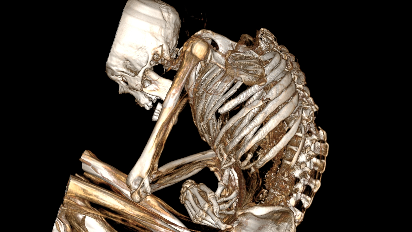 1,100-year-old mummy found in Chile died of extensive injuries when a turquoise mine caved in, CT scans reveal
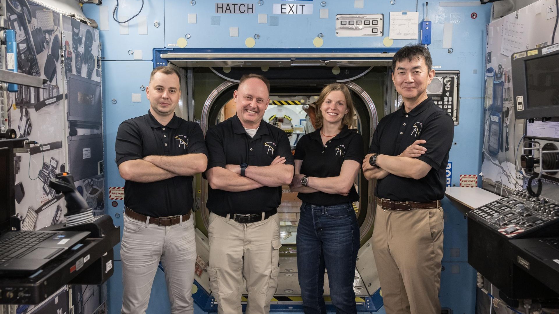 NASA announces unprecedented return of sick ISS astronaut and crew
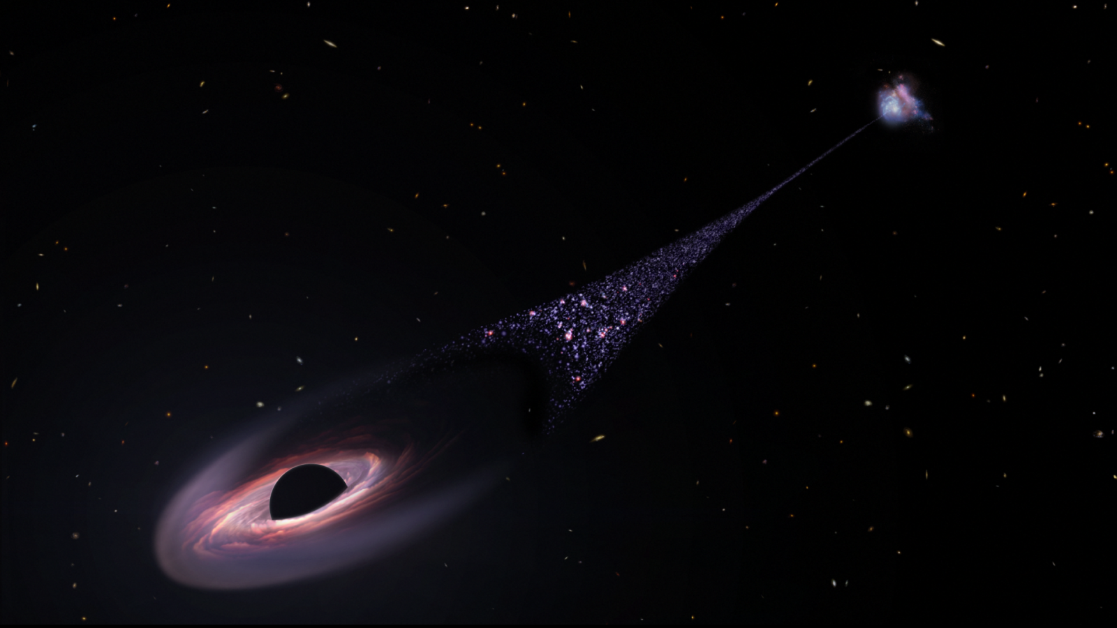 James Webb telescope confirms a supermassive black hole running away from its host galaxy at 2 million mph, researchers say
LATEST ARTICLES
James Webb telescope confirms a supermassive black hole running away from its host galaxy at 2 million mph, researchers say
LATEST ARTICLES- 1China's 'artificial sun' reactor shatters major fusion limit — a step closer to near-limitless clean energy
- 2Homo erectus wasn't the first human species to leave Africa 1.8 million years ago, fossils suggest
- 3Giant sunspot that triggered recent solar 'superstorm' shot out nearly 1,000 flares and a secret X-rated explosion, record-breaking study reveals
- 4The Waorani capture an Anaconda in Bameno, Ecuador.
- 5Tiny bump on 7 million-year-old fossil suggests ancient ape walked upright — and might even be a human ancestor